The next generation of Neuropixels brings us closer to understanding the brain
Neuropixels 2.0 packs over 5000 recording sites on a tiny brain implant enabling neuron recordings across brain regions and circuits. This will boost our knowledge of the brain.

In short
Neuroprobes are thin brain implants that can record brain activity
The new Neuropixels 2.0 probe increases the number of recording sites drastically to over 5000 while keeping the implantable area small
This technical challenge was made possible by advanced nano- and micro-processing technologies that integrate all the electronics on the neuroprobe itself
Neuropixels 2.0 enable brain-wide recordings which will boost our knowledge of how the brain works and how brain diseases originate
This article has been previously published in EETimes
Mapping the brain
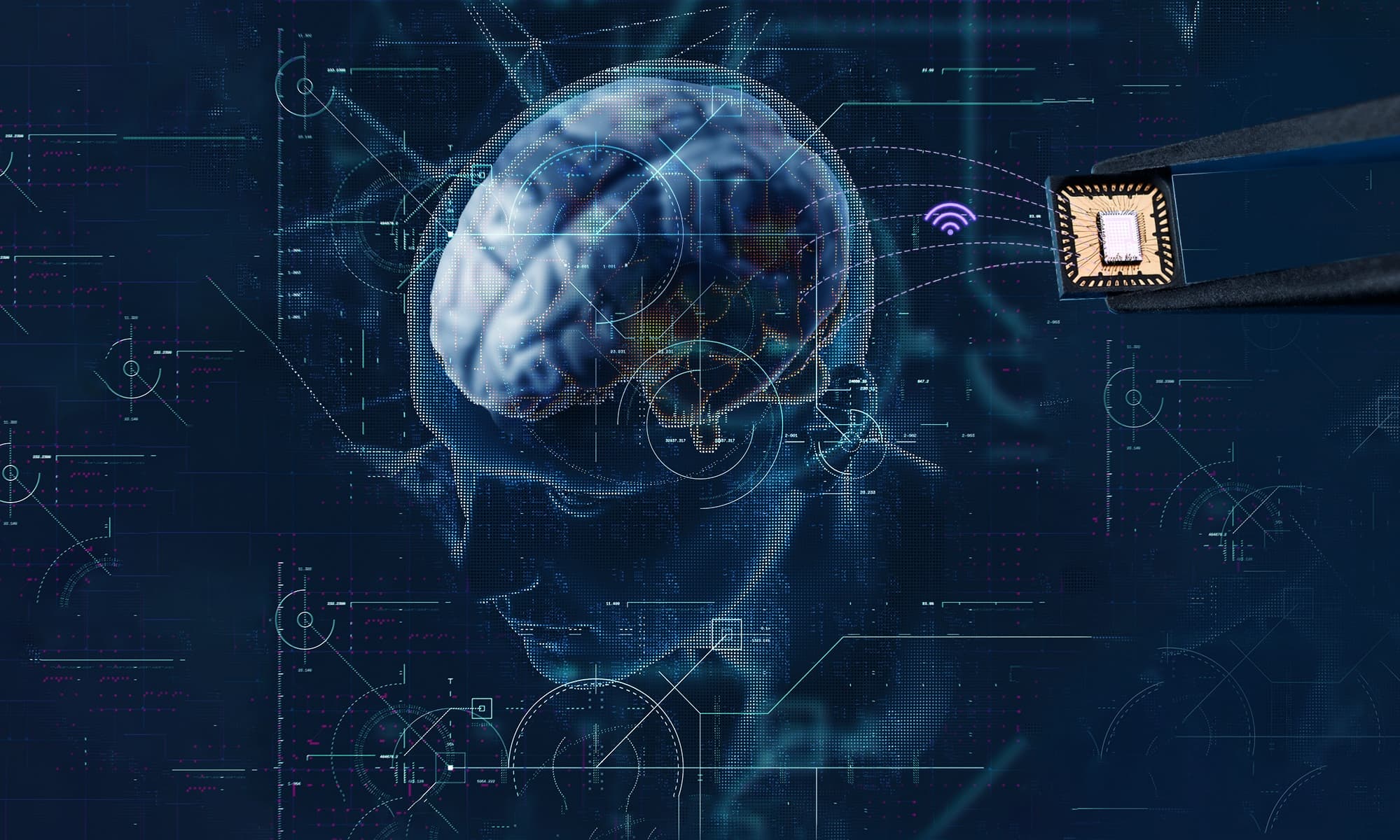
The story about studying the brain is also a story about designing technology to do so. One of the most successful neuroscientific devices of the last decades are neuroprobes or tiny needle-shaped brain implants that can pick up signals from individual neurons. Recording brain activity provides a unique look into how neurons communicate in intricate circuits to process information and control behavior. Large-scale recordings are ultimately needed to understand how the brain works and to develop more advanced brain-machine interfaces.
The number of recording sites or electrodes on the device started with just one on the tip of a single wire but increased to several tens with the introduction of silicon micro- and nanofabrication techniques. The first Neuropixels probes made the neuroscientific community change gears with close to a thousand electrodes on the implantable part, the shank. The new generation of Neuropixels (referred to as Neuropixels 2.0) now steps up to the plate with over five thousand electrodes distributed over four shanks, yielding unprecedented resolution for mapping brain activity.
How to connect thousands of electrodes
The challenge for such high-density devices is to connect each electrode to an external recording system while keeping the overall width of the shank as narrow as possible. The number of electrodes is therefore limited by the number of wires that can be accommodated in the shank. Increasing the width of the implantable part can lead to neuron damage and affect signal quality. To solve this, electronics were integrated on the probe itself using advanced CMOS-technology. This enabled multiplexing of the electrodes, so that the signals from several recording sites travel along the same cable.
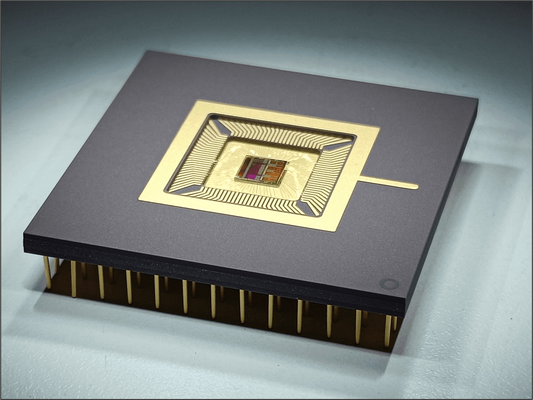
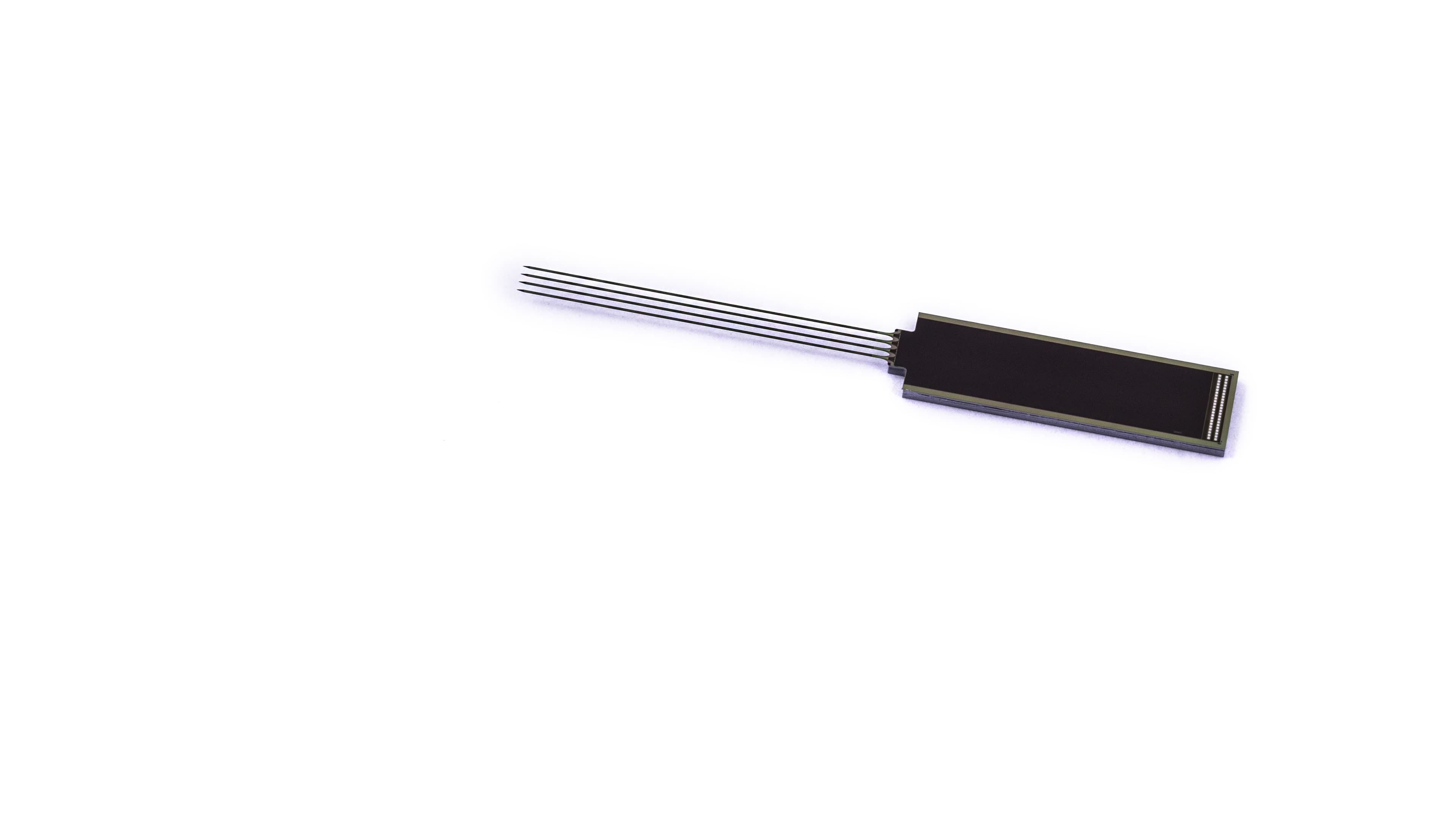
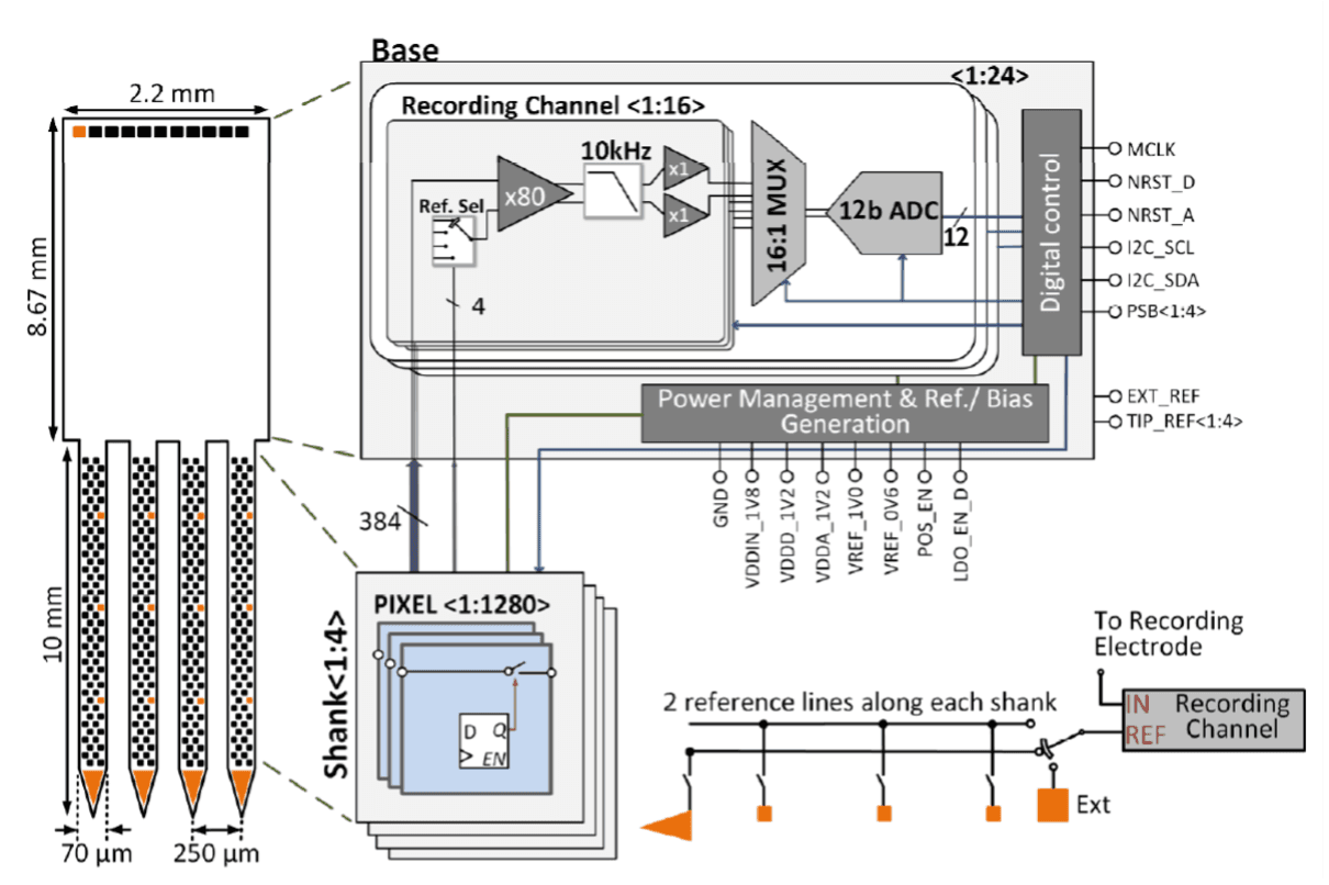
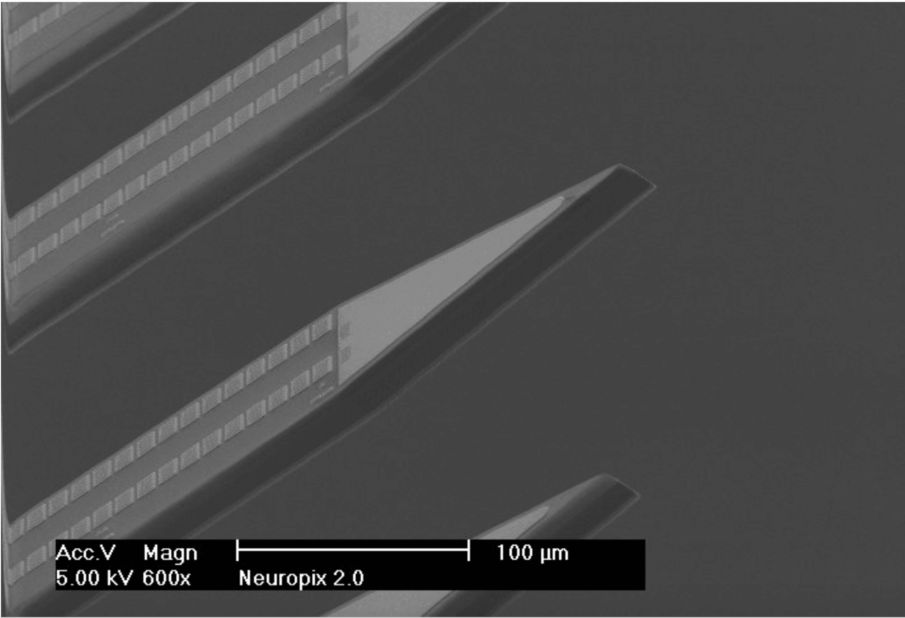
Neuropixels 2.0 thus packs 1,280 switchable electrodes or ‘pixels’ on a 70-by-24µm2 shank (5,120 for four shanks) with 384 interconnects. The electrodes are 12-by-12µm2 and integrate an analog switch and a 1-bit memory cell. The analog switches control which set of 384 electrodes are recorded simultaneously at any given time. This architecture makes it possible to select a pseudorandom combination of pixels along the shank. Moreover, the four shanks can densely sample activity from a 1-by-10mm2 plane perpendicular to the brain surface. Recording neural activity with such high coverage, spanning several brain regions and circuits simultaneously, is definitely a game-changer in the neuroscientific community.
Base electronics
The shank is monolithically integrated with a non-implantable part, the base, using 130nm CMOS processing technology. The base (8.67-by-2.2mm2) contains the electronics for multiplexing, amplification, digitization, and power management. The neuronal signals are first pre-processed on the probe base itself instead of using external equipment to ensure that no signal degradation occurs when the signals are transmitted out of the probe. Because the voltage signals are so small, they need to be amplified. The amplified analog signals are digitized to avoid noise and interference pick-up as they travel through the cable. To be able to record the full frequency band, a high-resolution 12-bit ADC (analog-to-digital converter) was integrated in the base. Finally, the ‘power management’ block was included in the probe to generate supply and reference voltages. The base electronics consume very low power (36.5mW) to minimize heating of brain tissue. At the same time, moving functionality onto the chip taps into the idea of system optimization with minimal components outside the probe, leading to a reduced size and weight of the overall system.
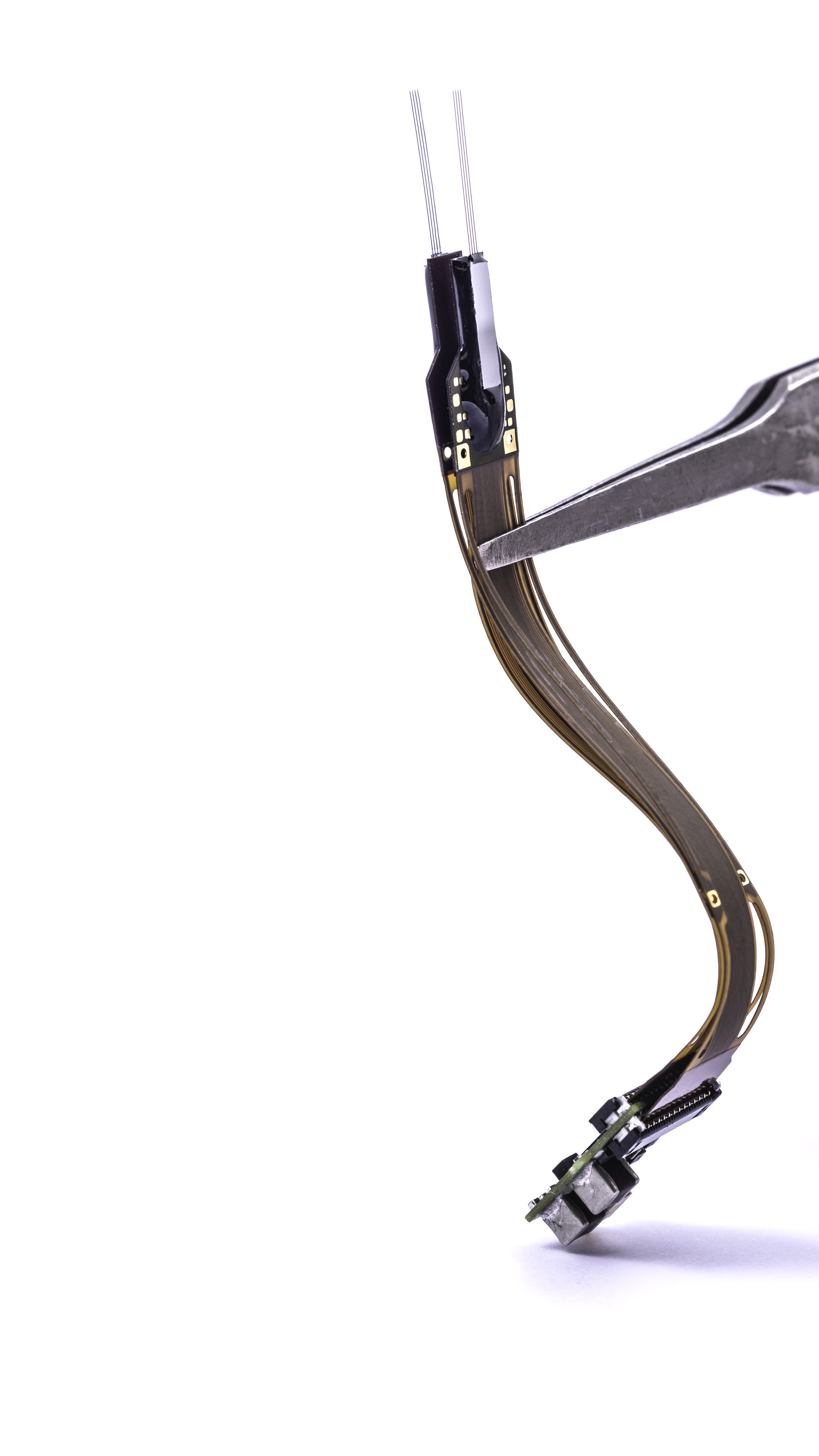
The digital output from all channels is subsequently transmitted over a 4-cm long flexible cable to an interface board (or headstage) that has a connection to a PXIe (peripheral component Interconnect (PCI) eXtension for instrumentation”; a standardized modular electronic instrumentation platform) card that acquires the data and sends it to a computer. The interface board of Neuropixels 2.0 was miniaturized to 10-by-14.3mm2. To make the system even more compact, it is possible to fit two probes onto one headstage with a combined weight of only 1.1g. If two four-shanks are used, the user has access to 10,240 recording electrodes. Moreover, new implantation hardware enables recovery and re-use of the implanted probes.
Long-term recordings
One of the main challenges for neuroscientists is to record from the same neurons stably for days and weeks to study the neural basis of processes that evolve over time such as learning or memory. Neurons frequently ‘drift’ making it difficult to track them based on their signal characteristics. With long-term recordings in mind, the electrode layout of Neuropixels 2.0 is linearized in two columns rather than staggered along the shank. This allows a new stabilization algorithm to lock onto a neuron’s unique signature. The algorithm takes advantage of the redundancy of the signal on closely spaced neighboring electrodes (pitch 15µm) to determine if it’s the same neuron.
The electrodes were fabricated out of the relatively new material TiN (titanium nitride) that proved to be very stable for long-term recordings. The electrodes have an impedance of 148 ± 8 kΩ at 1kHz. The system achieves a mean input-referred noise of 7.44 μVrms in the action-potential band (single neuron activity) and 7.65 μVrms in the local-field-potential band (summed neuron activity).
Decoding behavior
These unique features of Neuropixels 2.0 allows recordings of hundreds of identifiable neurons for over weeks to months. This new scale of recordings heralds a new era in brain research where neuroscientists can now investigate large populations of neurons spanning multiple brain regions. It enables the study of the dynamics in connected circuits that underlie behavior and thought.
Recent brain-wide studies with the first generation of Neuropixels probe explored how behavior is encoded in the brain. Thanks to this new technology, flows of neuronal activity can be visualized across the brain when an animal senses, or performs a task. The results show that complex behavior is generated through the interaction of groups of neurons spread out over the brain. Moreover, brain activity is driven by a mix of task-related behavior, spontaneous behavior (e.g. whisking, sniffing) and motivational state (e.g. being thirsty). Especially this last finding is surprising as until now most studies focused on associating neural activity with an external variable such as a task or an induced behavior, while unrelated aspects of the recording are classified as ‘noise’. The recent studies use large-scale recordings to show that the latent activity that is picked up by the probes brain-wide corresponds to ongoing behavior from the animal.
These first studies show how large-scale recording tools can propel our knowledge of the brain forward. With even more electrodes and a smaller size, Neuropixels 2.0 will be able to capture more information simultaneously, which will, ultimately, bring us closer to understanding how the brain works and how brain diseases arise.
The Neuropixels 2.0 consortium is led by Tim Harris at the Howard Hughes Medical Institute’s Janelia Research Campus, and includes funding from and scientists at University College London (UCL), at the Norwegian University for Science and Technology (NTNU) in Trondheim, Norway, at Neuroelectronics Research Flanders (NERF) in Leuven, Belgium, and at the Champalimaud Centre for the Unknown in Lisbon, Portugal.
Want to know more
For information about the probes, ordering, training, and the latest on the next generation, visit www.neuropixels.org.
More information about imec’s neural probe program can be found on their website.
The research information about the Neuropixels probes has been published in following papers, which can be requested through this form.
- Jun, J., Steinmetz, N., Siegle, J. et al. Fully integrated silicon probes for high-density recording of neural activity. Nature 551, 232–236 (2017). DOI: 10.1038/nature24636.
- Steinmetz N.A. et al. Neuropixels 2.0: A miniaturized high-density probe for stable, long-term brain recordings. Science 372, eabf4588 (2021). DOI: 10.1126/science.abf4588.
- Wang S., Garakoui S.K., Chun H., Salinas D.G., Sijbers W., Putzeys J., Martens E., Craninckx J., Van Helleputte N., Lopez C.M. A Compact Quad-Shank CMOS Neural Probe With 5,120 Addressable Recording Sites and 384 Fully Differential Parallel Channels. IEEE Trans Biomed Circuits Syst. 2019 Dec;13(6):1625-1634. DOI: 10.1109/TBCAS.2019.2942450. Epub 2019 Sep 19. PMID: 31545741.
- Mora Lopez C., Putzeys J., Raducanu B.C., Ballini M., Wang S., Andrei A., Rochus V., Vandebriel R, Severi S., Van Hoof C., Musa S., Van Helleputte N., Yazicioglu R.F., Mitra S. A Neural Probe With Up to 966 Electrodes and Up to 384 Configurable Channels in 0.13 µm SOI CMOS. IEEE Trans Biomed Circuits Syst. 2017 Jun;11(3):510-522. DOI: 10.1109/TBCAS.2016.2646901. Epub 2017 May 19. PMID: 28422663.
- Allen WE, Chen MZ, Pichamoorthy N, Tien RH, Pachitariu M, Luo L, Deisseroth K. Thirst regulates motivated behavior through modulation of brainwide neural population dynamics. Science 2019 Apr 19;364(6437):253.
- Stringer C, Pachitariu M, Steinmetz N, Reddy CB, Carandini M, Harris KD. Spontaneous behaviors drive multidimensional, brainwide activity. Science. 2019 Apr 19;364(6437):255.
- Musall S, Kaufman MT, Juavinett AL, Gluf S, Churchland AK. Single-trial neural dynamics are dominated by richly varied movements. Nat Neurosci. 2019 Oct;22(10):1677-1686.
Authors

Carolina Mora Lopez
Carolina Mora Lopez received the Ph.D. degree in electrical engineering from KU Leuven, Leuven, Belgium, in 2012, in collaboration with imec, Leuven. From 2012 to 2018, she worked as a Researcher and an Analog Designer with imec focused on interfaces for neural-sensing applications. She is currently the Scientific Director and the Team Leader of the circuits for neural interfaces team with imec. Her research interests include analog and mixed-signal circuit design for sensors, bioelectronics, and neural interfaces. Dr. Mora Lopez is an IEEE SSCS Distinguish Lecturer and serves on the Technical Program Committee of the VLSI Circuits Symposium, International Solid-State Circuits Conference (ISSCC), and ESSCIRC conferences.